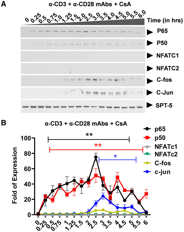
Fig. 3.
Western blot analysis showing specific inhibition of nuclear NFAT induction following TCR activation. (A) Clone 2D10 cells were pretreated with calcium-calcineurin inhibitor cyclosporine A (CsA) for one hour prior to the time-course activation experiment. Nuclear fractions were used to determine specific inhibition of NFAT, AP-1, and both AP-1 and NFAT nuclear induction using western blot analysis. Spt5 was used as the loading control. Note that the results presented here are a representation of immunoblot experiments performed in triplicates. (B) Densitometry analysis of relative nuclear NF-κB (p65 and p50), NFAT (c1 and c2), and AP-1 (c-Fos and c-Jun) protein levels in (A) above. Error bars represent the mean ± SD of three independent and separate experiments. The P value of statistical significance was set as P < 0.05 (*) or 0.01 (**).

