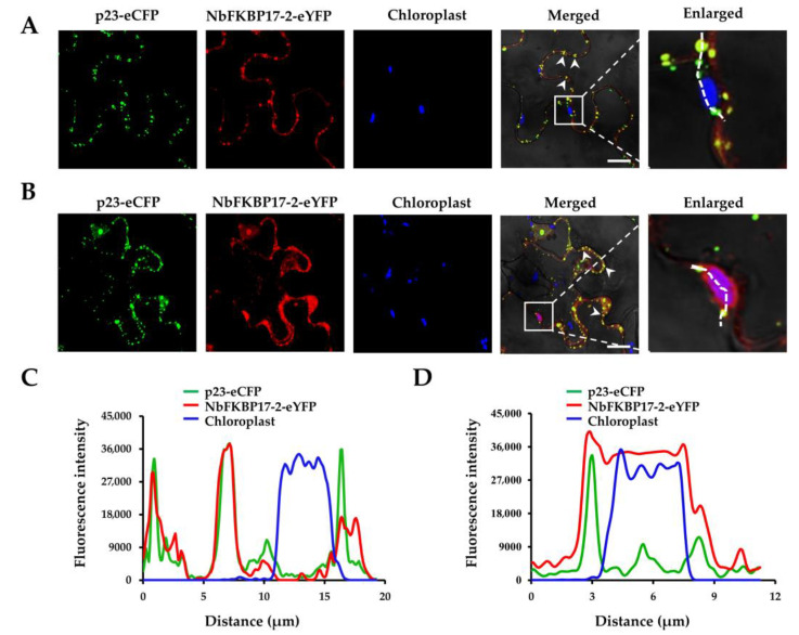
Figure 3.
Subcellular co-localization analyses of p23-eCFP and NbFKBP17-2-eYFP in Nicotiana benthamiana epidermal cells. (A,B) represented two different visual fields showing the co-localization of p23 and NbFKBP17-2 at plasmodesmata (PD) (A) and partial fluorescent signals of NbFKBP17-2 on chloroplasts (B), respectively. The white boxes indicated areas showing a magnified view. Arrows point to PD. The blue signals represented the chlorophyll autofluorescence in chloroplasts. Confocal images were taken at 48 hpi. Bars, 20 μm. (C,D) showed overlapping fluorescence spectra of eCFP, eYFP and chloroplast signals along the white dots depicted in (A,B), respectively.

