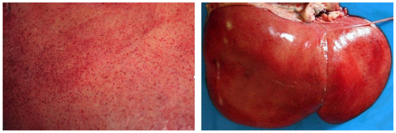
Figure 1.
Macroscopic pathology of the liver in sheep naturally infected with RVFV. (A) Innumerable petechia on the parietal surface of the liver of an adult sheep. (B) Liver of a new-born lamb with extensive necrosis and pinpoint subcapsular petechia giving the liver a pale yellow to red mottled appearance.

