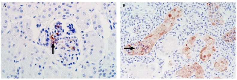
Figure 7.
Immunolabelling for RVFV in the kidney of sheep (NovaRed IHC as detailed in Figure 5). (A) In this specimen from a young lamb, viral antigen is prominent in Lacis cells in the glomerulus (arrow). Other cells, morphologically consistent with endothelial cells also labelled in the glomerulus. Cells in the macula densa are not labelled, mag 400×. (B) Adult sheep with immunolabelling of necrotic renal tubular epithelial cells (arrow) in the renal cortex, mag 600×.

