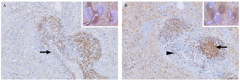
Figure 9.
Spleen of an RVFV-infected adult sheep with sequential sections immunolabelled for T and B lymphocytes respectively (polyclonal rabbit anti-CD3 and anti-CD20 antibodies, micro-polymer detection system, DAB chromogen and haematoxylin counterstain). (A) Labelling with the anti-CD3 antibody shows a marked loss of T lymphocytes. Only scattered T lymphocytes remain in the red pulp and there is severe depletion of the periarteriolar lymph sheath (arrow), mag 100×. Inset: Spleen from a healthy control sheep showing a normal distribution of abundant T lymphocytes, mag 100×. (B) Labelling with the anti-CD 20 antibody shows necrotic debris in the germinal centre (arrow) with a few residual B lymphocytes in the marginal zones (arrowhead) of the periarteriolar lymph sheath and in the red pulp, mag 100×. Inset: Spleen from a healthy sheep with multiple lymphoid follicles that contain many B lymphocytes, mag 100×.

