Figure 2.
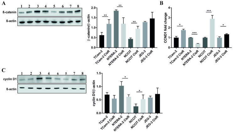
NTERA-2 CisR cells exhibited decreased levels of β-catenin and cyclin D1 compared to parental cells. (A) The western blot analysis of β-catenin showed decreased level of this protein in NTERA-2 CisR cells. Other GCT cell lines had increased expression of β-catenin on the protein level what was confirmed also by densitometric analysis. (B) Only chemoresistant NTERA-2 CisR cells exhibited the decrease in CCND1 expression as demonstrated by qPCR. (C) Western blot and densitometric analysis confirmed significantly decreased expression of cyclin D1 also on the protein level. 1. TCam-2; 2. TCam-2 CisR; 3. NTERA-2; 4. NTERA-2 CisR; 5. NCCIT; 6. NCCIT CisR; 7. JEG-3; 8. JEG-3 CisR. β-actin was used as an internal loading control. * p < 0.05, ** p < 0.01, *** p < 0.001.

