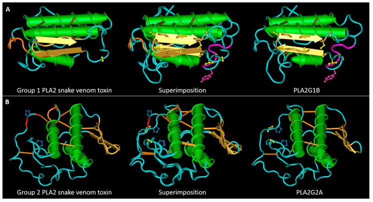
Figure 4.
Main SLiMs that differentiates snake venom PLA2s from their mammalian homologues. (A) 3D structures of the snake venom neuro-myotoxin notexin, of human PLA2G1B and their superimposition. The S/T residues (represented in yellow) in the central region of PLA2G1B, belong to four superimposed motifs phosphorylable by GSK3. The tyrosine is represented in magenta form, together with two residues of the pancreatic loop. The motif of interaction with I-BARREL proteins: the loop colored in orange in the group I toxin contains, in numerous cases, an SH2 and PTB binding sites. (B) 3D structures of the snake venom myotoxin bothropstoxin-I, of human PLA2G2A and their superimposition. The S/T residue evidenced in yellow in PLA2G2A, when phosphorylated, allows isomerization of the adjacent proline by Pin1. The loop following the second α-helix in the toxin structure, colored in orange, contains a PKA phosphorylation site and an SH2 binding site. The third and last C-terminal amino acid of the toxins (colored in red) forms a PDZ binding motif. The PDB files used for this figure are the same as those described for Figure 1.

