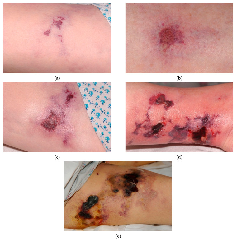
Figure 1.
Examples of typical calciphylaxis wound presentations. As shown in the images, milder calciphylaxis wounds (a–c) typically have color changes in the surrounding skin and relatively low exudate, without evidence of infection. More severe, advanced calciphylaxis wounds (d,e) develop ulceration and necrotic tissue with eschar obscuring the actual depth of the wound, as well as edema and erythema suggesting inflammation. When palpated, there is induration adjacent to the necrotic tissue in the more severe wounds.

