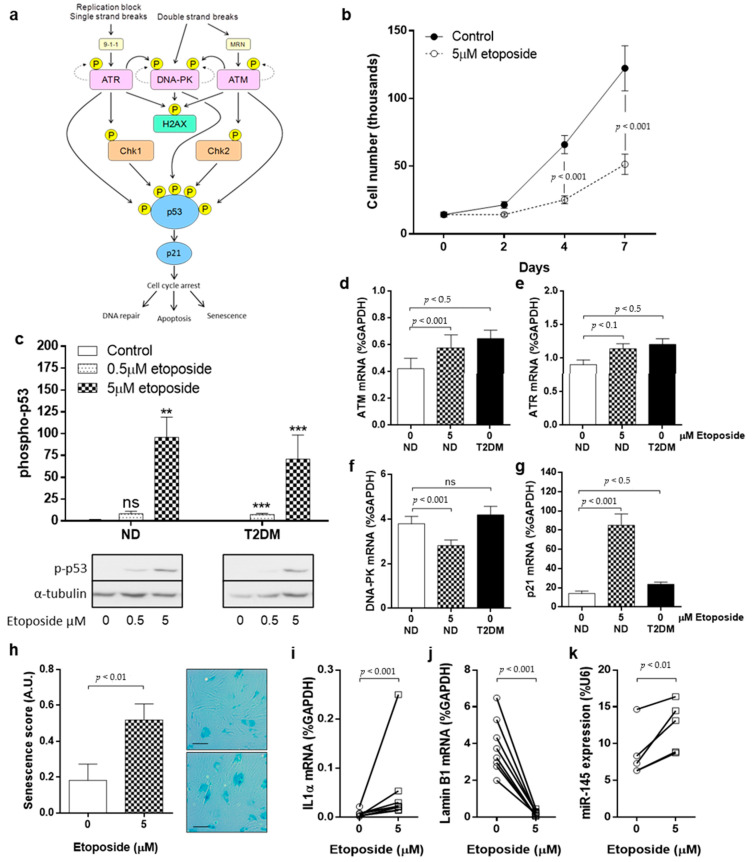
Figure 2.
DNA damage drives a senescent SMC phenotype. (a) The DNA damage signalling pathway leading to phosphorylation of p53, and subsequent downstream effects. (b) SMC were treated with DNA-damaging agent etoposide (5 μM) or vehicle control (DMSO) for up to 7 days and proliferation quantified by cell counting (n = 7). (c) The impact of etoposide on p53 phosphorylation was monitored using Western blotting after 24 h (n = 4). (d) The effect of DNA damage on apical kinase expression was explored in ND-SMC that were treated with 5 μM etoposide for 24 h. The expression of ATM, (e) ATR, (f) DNA-PK and (g) p21 was quantified using RT-PCR. The expression of these kinases was also measured in untreated T2DM-SMC to determine whether DNA damage per se could mimic a T2DM-SMC phenotype (all n = 8). (h) Cells were treated with etoposide for 24 h and then placed into FGM for 72 h. Cells were stained with SA-β-galactosidase (scale bar = 100 μm). (i) The influence of etoposide on expression of IL-1α, (j) LMNB1 (both n = 8) and (k) miRNA-145 expression (n = 6–8) was quantified using RT-PCR.

