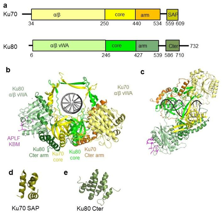
Figure 2.

Structure of the human Ku70/80 heterodimer. (a) Human Ku70 and Ku80 have similar organizations. They present some sequence variabilities in their C-terminal region. (b,c) Crystal structure of Ku70/80 complexed with a hairpin DNA and the Ku binding motif of APLF (magenta) [26]. Ku is colored in the same way as that in (a). The right view shows the top view of the complex. The DNA is a hairpin DNA used to limit Ku movement on the DNA for crystallization. (d) Structure of the SAP domain of Ku70 solved by NMR [48]. (e) C-terminal domain of Ku80 solved by NMR [49,50].
