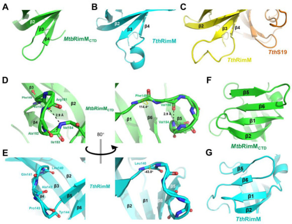
Figure 3.

Structural comparison between MtbRimMCTD and RimM from T. thermophilus HB8 (TthRimM) represented in cartoon. (A–C) β3-β4 loop of MtbRimMCTD (A), free TthRimM (B), S19-complexed TthRimM (C). (D,E) β4-β5 loop of MtbRimMCTD (D) and free TthRimM (E). The dihedral angle ψ of F149 in (D) and L140 in (E) are identified. The length of the hydrogen bond (V154)N-H…O(V150) is also depicted in (D), where backbone oxygen or nitrogen atoms are shown as red and blue sticks, respectively. Hydrogen atoms, if applicable, are hidden. (F,G) β5 and β6 strands of MtbRimMCTD (F) and free TthRimM (G).
