Figure 4.
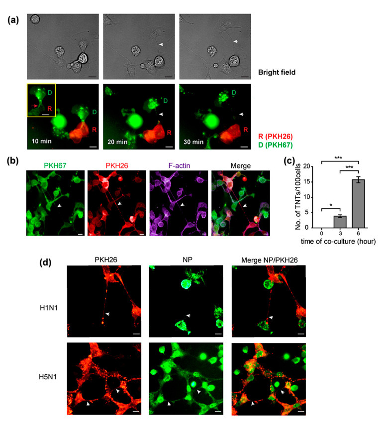
Tunneling nanotubes (TNTs) are formed between donor and recipient cells after trogocytosis. MDCK cells were infected with H5N1 at an MOI of 1 for 8 h. Infected MDCK cells labeled with PKH67 were used as donor cells. Donor and PKH26-labeled recipient cells were directly co-cultured. Cells were then imaged every 10 min using live-cell microscopy. Live cell images showed the TNT formation (indicated as white arrow) between the infected donor and recipient cells after trogocytosis ((a), middle and right panels). The red box shows the PKH67-labeled membrane of donor cells transferred to recipient cells (trogocytosis) indicated as a red arrow ((a), left panel). The co-cultures were fixed and stained for F-actin. (b) The fluorescence images showed the presence of PKH67, PKH26, and F-actin on the TNT indicated as an arrow. (c) The TNTs were then quantified at the indicated time point. Co-culture assays of H5N1 and H1N1 infection were performed following Figure 1a. (d) The presence of tunneling nanotubes (arrow) was demonstrated in both H5N1 and H1N1-infected co-cultures. (d) NP existed in the TNT as indicated by the arrow in the NP panel. The data are presented as the mean ± SEM of three independent experiments. Statistical analysis was performed using one-way ANOVA * p < 0.05, *** p < 0.001. Scale bars: 10 µm.

