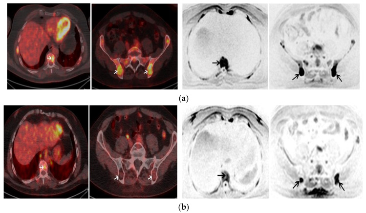
Figure 4.
A 55-year-old male with IgA kappa MM. (a) Axial images of baseline 18-FDG PET/CT (left panel) shows multiple focal lesions (FL) with high uptake including one FL involving T9 and 2 FL involving the pelvis (white arrows). Axial images of baseline WB-DW-MRI (left panel) shows FL with restricted diffusion (b = 800 s/mm2) involving also T9 and the pelvic bone with an ADC of 920 (black arrows); (b) Positive post-ASCT 18-FDG PET/CT (left panel) shows a persistent uptake of T9 (FL SUVmax > liver uptake) and the regression of uptakes within FL involving the pelvis. Positive post-ASCT WB-DW-MRI (right panel) shows a regression of the restricted diffusion in T9 but the persistence of two FL in the pelvis with restricted diffusion and ≤25% increase of the ADC values (ADC = 1000). Patient was in complete remission after ASCT but relapsed 16 months after ASCT.

