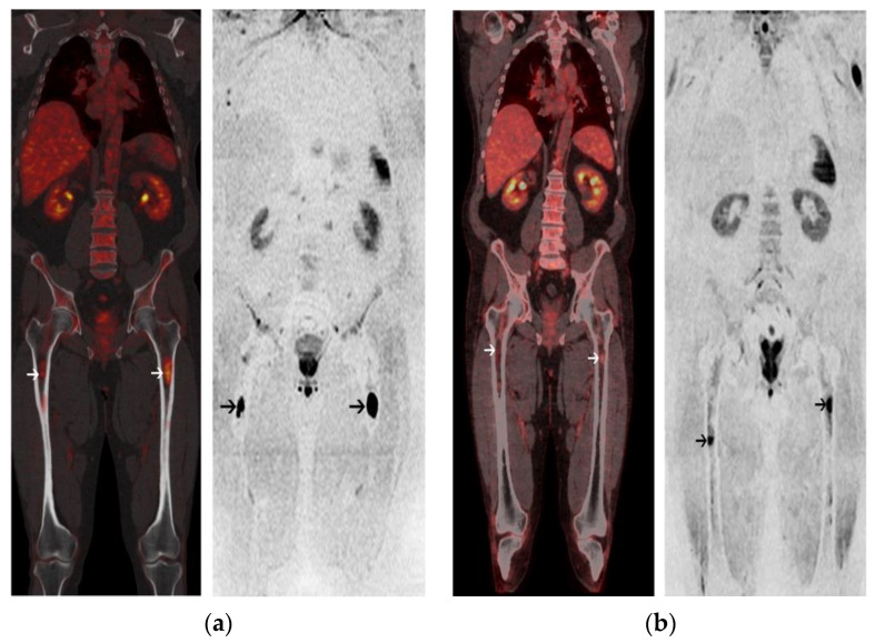
Figure 5.
A 63-year-old male with Kappa Light Chain MM. (a) Coronal image of 18-FDG PET/CT at baseline (left panel) shows 2 focal lesions (FL) within the femurs (white arrows). Corresponding coronal image of baseline WB-DW-MRI (right panel) shows 2 FL with restricted diffusion within the femurs; (b) Post-ASCT 18-FDG PET/CT is negative and coronal image (left panel) shows a regression of uptake of the femoral FLs (FL SUVmax < liver uptake) (white arrows). Post-ASCT WB-DW-MRI shows a regression of the extent of the femoral FL (black arrows) associated with a decrease of the diffusion restriction and a significant increase of the ADC value from baseline (>40%). Patient was classified as low-risk by both imaging modalities and is in complete remission after 33 months of follow-up.

