Figure 9.
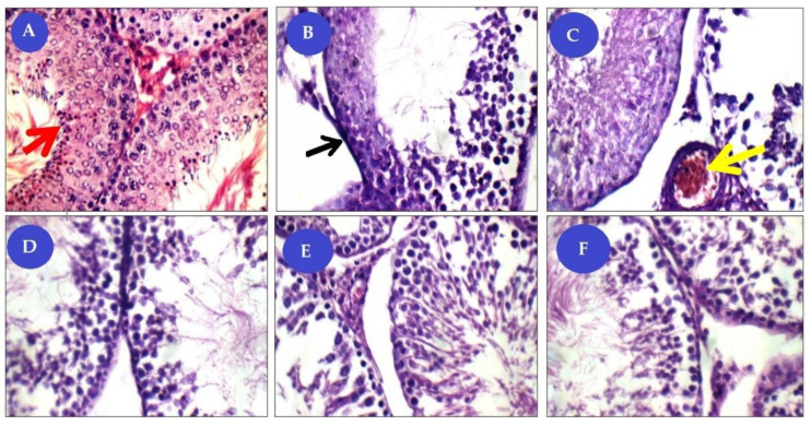
Panel of histological (H&E ×400) pictures showing: (A) control group (NG) seminiferous tubules (ST) with average BM (basement membrane), spermatogonia, primary spermatocyte, and many spermatozoa (red arrows); (B,C) Cisplatin-treated group (CG) with tubules have detached and thick BM (black arrow), marked reduction of germinal lining with few sperms, and interstitial blood vessels congested (yellow arrow). (D) Cisplatin+ mesenchymal stem cells treated group (CMG) tubules with mildly thick BM, mild reduction of germinal lining with moderate number of sperms. (E) Cisplatin+ mesenchymal stem cells+ beetroot extract treated group (CBG) tubules with average BM, average germinal lining up to full spermatogenesis, and average interstitium with average Leydig cells. (F) Cisplatin+ mesenchymal stem cells+ beetroot extract treated group (CMBG) tubules with mildly thick BM, average germinal lining up to full spermatogenesis, and average interstitium with average Leydig cells.

