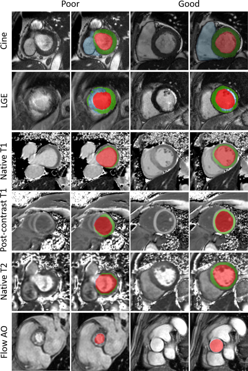
Fig. 4.
Results example of left cavity (red), left myocardium (green), right cavity (blue), scar tissue (blue), and aorta (red) segmentations obtained using the pipeline. Good, and poor results are shown for cine, LGE, T1, post-contrast T1, T2, and aortic flow images. The pipeline faces multiple challenges: the basal slice with its variability, and noise (CINE, T1, and T2); clouded and undefined boundaries (the myocardium on LGE images); artifacts compromising the shape of anatomical structures (post-contrast T1); and irregularities. Flow AO = aortic flow

