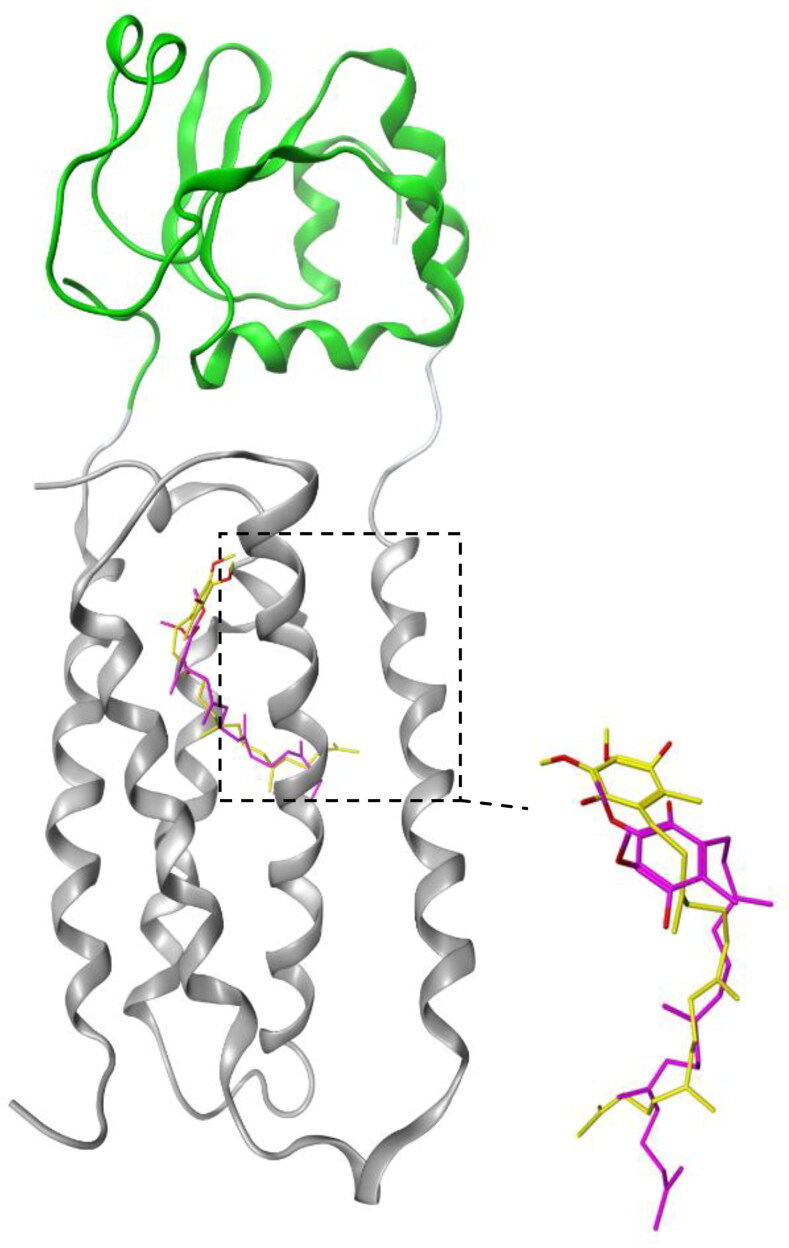
Figure 3.
Superposition of the docked conformer of quinone with the bacterial VKOR. The bacterial VKOR and Trx-like domain are shown in gray and green ribbons, respectively. The dotted square contains the quinone binding site. Yellow and magnetic sticks represent the quinone molecules in the x-ray crystal structure of VKOR obtained from experimental crystallography and docking analysis, respectively.

