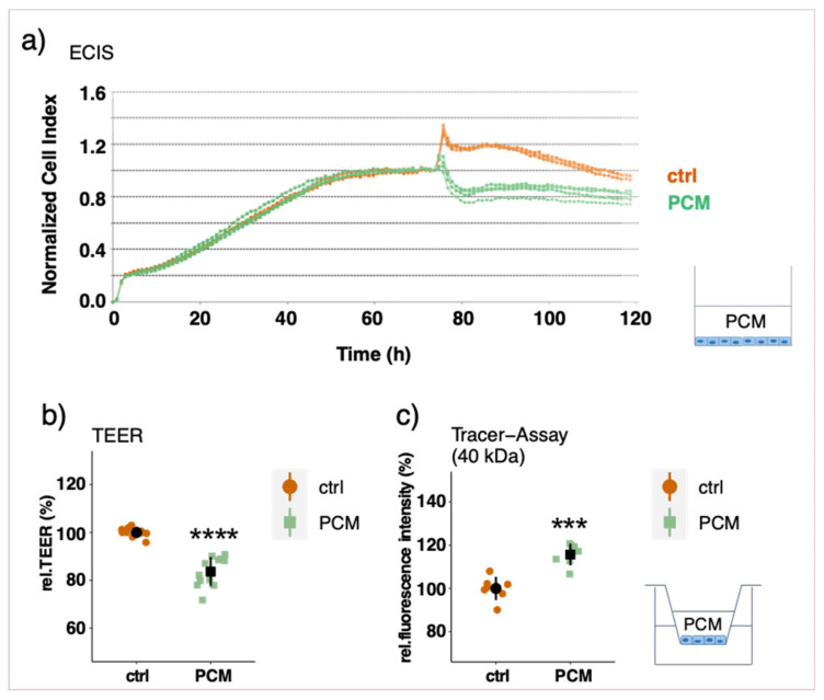
Figure 5.
Measurements of endothelial barrier function after apical PCM treatment of an EC monolayer. ECs were cultured in eplates (a) or on transwell inserts (b,c). After a stable barrier function was established (6–7 days), PCM was added to the apical side for 24 h. Representative real-time ECIS measurements with the xCELLigence system (a), relative trans-endothelial electric resistance (TEER) measurements with a CellZcope instrument (b) and macromolecular tracer assays with FITC-labeled dextran of 40 kDa (representative also for 4 kDa) (c). Experiments were performed at least three times in triplicates and data represent mean ± sd. *** p < 0.001, **** p < 0.0001.

