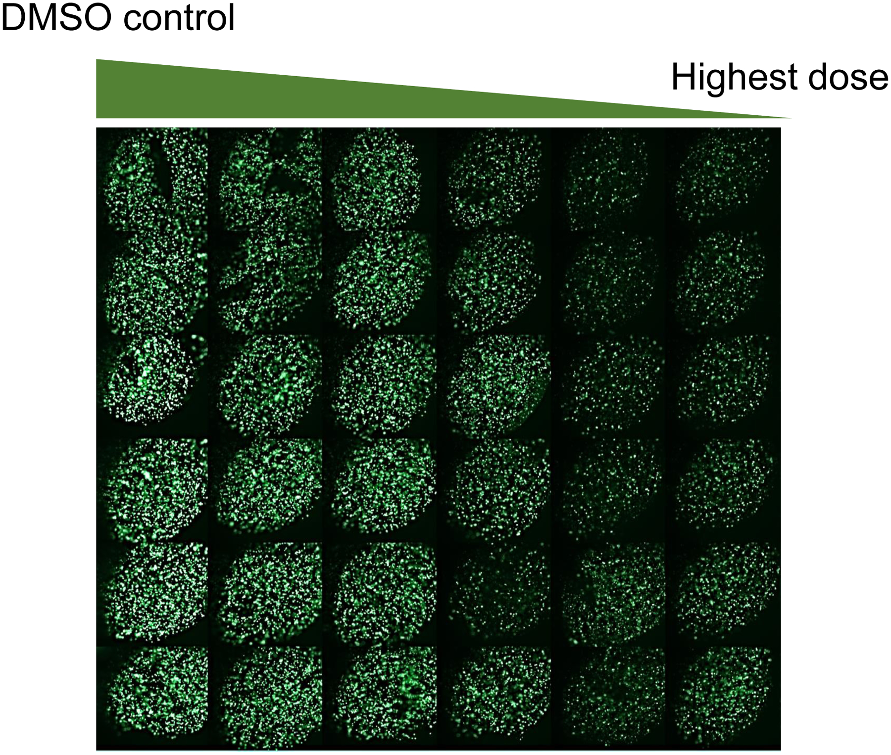
Figure 4.

Fluorescent images of 3D-cultured ReNcell VM cells on the 384-pillar plate, obtained by exposing the cells to DMSO (vehicle control) and 0.08 – 20 μM of topotecan for 24 hours with 6 replicates in each row, staining with 1 μM of calcein AM for 1 hour, and scanning with a green fluorescence filter of S+ Scanner.
