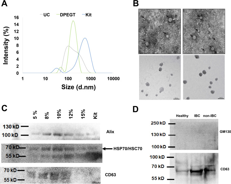
Fig 2. Characterization of enriched plasma sEVs of breast cancer patients.
The successful isolation of plasma sEVs was verified by different methods. (a) sEVs size distribution was determined by dynamic light scattering (DLS). (b) Transmission electron micrograph of negatively stained sEVs. Size bar equal 200 nm. (c) Western blot of the exosomal proteins markers; Alix, CD63 and HSP70/HSC70 using different concentrations of DPEGT (pooled, n = 12) in comparison to the miRCURY kit. (d) Western blot analysis of the exosomal proteins (e.g., CD63) and non-exosomal Golgi apparatus protein GM130.

