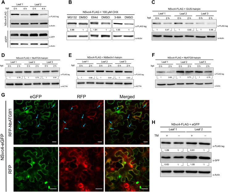
Fig 2. NSvc4 is unstable in plant cells and degraded through the autophagy pathway.
(A) In vivo protein stability assay of NSvc4. The leaves expressing NSvc4-FLAG or eGFP were treated with 100 μM CHX. Samples were harvested at 0 h and 4 h after treatment for western blotting. Actin was used as a loading control. (B) Protease inhibitors treatment of the leaves expressing NSvc4-FLAG. The leaves expressing NSvc4-FLAG were treated with 100 μM CHX together with MG132, E64d, 3MA, or DMSO, respectively. Six hours later, the leaves were harvested, and total protein was extracted for western blotting. Actin was used as a loading control. The relative levels of NSvc4-FLAG to Actin were calculated and normalized by setting values in DMSO treatment as 1.0. (C-F) In vivo protein degradation rate assay of NSvc4 upon silencing NbATG5, NbBeclin1 or NbATG8. The leaves expressing NSvc4-FLAG with GUS-hairpin (C), NbATG5-hairpin (D), NbBeclin1-hairpin (E) or NbATG8-hairpin (F) were treated with CHX. Samples were harvested at 0 h and 2 h after treatment for western blotting. Actin was used as a loading control. The relative levels of NSvc4-FLAG to Actin were calculated and normalized by setting values at 0 h as 1.0. (G) Confocal images of NSvc4-eGFP co-expressed with RFP-NbATG8f1. N. benthamiana leaves were infiltrated with agrobacteria carrying NSvc4-eGFP and RFP-NbATG8f1 or RFP. Arrows indicate colocalized eGFP and RFP fluorescence. Bars, 20 μm. (H) Western blotting analysis of NSvc4-FLAG after TM treatment in N. benthamiana leaves. NSvc4-FLAG and eGFP were co-expressed in N. benthamiana leaves by agroinfiltration. Then, the leaves were treated with TM at 42 hpi. Eighteen hours later, total protein was extracted for western blotting analysis. The relative levels of NSvc4-FLAG to Actin were calculated and normalized by setting values in DMSO control as 1.0.

