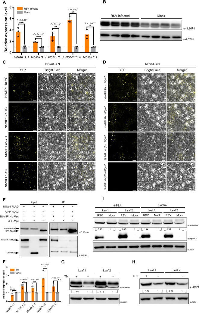
Fig 3. NbMIP1 family proteins are upregulated by RSV-induced UPR and interact with NSvc4.
(A) RT-qPCR analysis of NbMIP1 family expression level in RSV-infected or healthy N. benthamiana. NbActin was used as an internal reference in relative quantification. The values represent the means of the expression levels ± standard deviations (SD) relative to the mock plants (n = 3 biological replicates). Student’s t-test was performed, and asterisks denote significant differences between RSV-infected and mock plants (two-sided, *P < 0.05, **P < 0.01, ***P < 0.001). (B) Western blotting analysis of NbMIP1 family proteins in RSV-infected and mock plants at 12 dpi. Total protein was extracted for western blotting. Actin was used as a loading control. (C and D) BiFC assay of NSvc4 with NbMIP1.4b and its truncated versions. Leaves were infiltrated with agrobacteria carrying NSvc4 fused to the N-terminal part of YFP, and NbMIP1.4b or its truncated versions were fused to the C-terminal part of YFP. Samples were observed by laser confocal microscopy at 48 hpi. Bars, 50 μm. (E) Co-immunoprecipitation assay of NSvc4 with NbMIP1.4b. NSvc4-FLAG or GFP-FLAG were co-expressed with NbMIP1.4b-Myc or GFP-Myc in N. benthamiana leaves. 48 hours later, the leaf protein extracts were incubated with FLAG magnetic beads. Samples before (input) and after immunoprecipitation (IP) were analyzed by western blotting using anti-FLAG or anti-Myc antibodies. (F) RT-qPCR analysis of NbMIP1s expression levels after DTT treatment. Each half of a leaf was treated with DTT or ddH2O, respectively. The total RNA was extracted at 18 hpt. NbActin was used as an internal reference in relative quantification. The relative expression levels were normalized by setting the value of control as 1.0 in each leaf (n = 3 biological replicates). Then, data were analyzed by Student’s t-test, and asterisks denote significant differences between DTT-treated and control halves (two-sided, *P < 0.05, **P < 0.01, n.s., not significant). (G and H) Western blotting analysis of NbMIP1.4b in N. benthamiana leaves treated with TM (G) or DTT (H). The samples in Fig 1B and 1C were used for western blotting to detect NbMIP1s. The relative levels of NbMIP1s to Actin were calculated and normalized by setting values in control halves as 1.0. (I) Samples in Fig 1D were analyzed by western blotting using antibody against NbMIP1s. Actin was served as loading controls. RSV CP indicated virus infection.

