. 2021 Jan 25;2020:1100–1109.

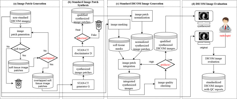
Figure 2: STAN-CT DICOM-to-DICOM image standardization framework. (a) Soft tissue image patches are generated from the input DICOM files, ready for image standardization and normalization. (b) For all the soft tissue image patches, new image patches are synthesized using STAN-CT generator. The quality of the new image patches is checked using STAN-CT discriminator. (c) All the synthesized soft-tissue image patches are integrated and are filtered by a soft tissue image mask generated using the input DICOM image. DICOM image quality is checked by examining box artifacts and empty pixels. (d) The synthesized and the original non-standard DICOM image files will be viewed side-by-side by radiologists using a PACS reading workstation. The radiologists’ reports will be used to further evaluate the quality of the standardized CT images. Meanwhile, image texture features will be extracted for automatic performance evaluation.

Figure 2: