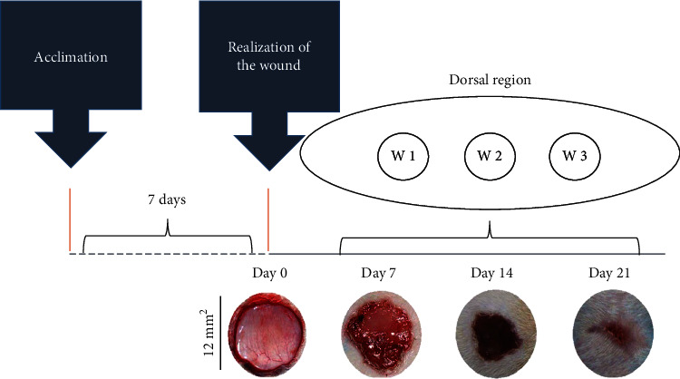
Figure 1.

Representation of the experimental model of wound healing by secondary intention and time-dependent evolution of wound closure. The top image shows the distribution of the three excisional wounds in the back of the animal. The general appearance of wound closure from the initial wound (day 0) is represented by photographs. W1 (day 7), W2 (day 14), and W3 (day 21); macroscopic aspect of the wounds observed every 7 days. The wound areas were calculated on days 0, 7, 14, and 21 (mean ± SD), based on the digitized images.
