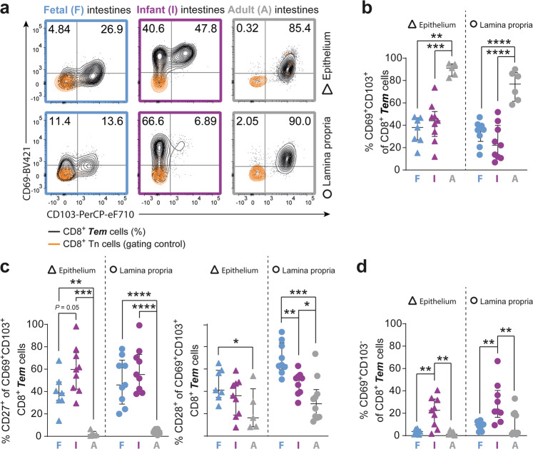
Fig. 2. Tissue-resident CD69+CD103+ CD8+ T cells increase after infancy.
a Representative flow cytometric plots of CD69 versus CD103 on CD8+ Tn (CCR7+CD45RA+; orange; gating control) and CD8+ Tem (CCR7-; black; %) cells in fetal (F; blue), infant (I; purple), and adult (A; gray) intestinal tissues. b Frequencies (%) of CD69+CD103+ CD8+ Tem cells. c Frequencies (%) of CD27+ and CD28+ cells within CD69+CD103+ CD8+ Tem cells. d Frequencies (%) of CD69+CD103− CD8+ Tem cells. Error bars represent median percentage ± IQR. The figure represents intestinal epithelium (fetal n = 7; infant n = 9; adult n = 5) and lamina propria (fetal n = 9, infant n = 9, adult n = 10) tissues. *P < 0.05, **P < 0.01, ***P < 0.001, ****P < 0.0001, all Mann–Whitney U analyses.

