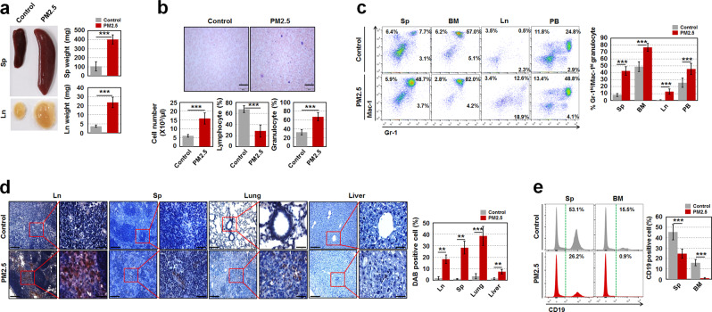
Fig. 2.
Maternal PM2.5-exposed offspring have the potential to develop a myeloproliferative disease. a Photographs of the Sp and Ln from control and maternal PM2.5-exposed offspring at 1 year of age and weights of those were measured (n = 4). b May–Grünwald–Giemsa stained blood smear (upper panels) and leukocyte number and the proportion of lymphocytes and granulocytes (lower panels) from the old offspring (n = 5). Scale bars are 200 µm. c Percentage of Gr-1+/Mac-1+ granulocytes in Sp, BM, Ln, and PB of the old offspring (n = 5). d Myeloperoxidase immunohistochemistry was conducted in hematopoietic (Ln and Sp) and nonhemtopoietic organs (lung and liver) of the old offspring to measure infiltrated blast cells to the organs. A representative result is shown (n = 5). Scale bars are 100 (left) and 20 (right) µm. DAB-positive cell intensity was measured by ImageJ-win64. e Percentage of B cells (CD19+ cells) in Sp and BM of the old offspring (n = 5). All data are presented as the means ± SD. **p < 0.01 and ***p < 0.001 vs. control, as determined by Student’s t tests

