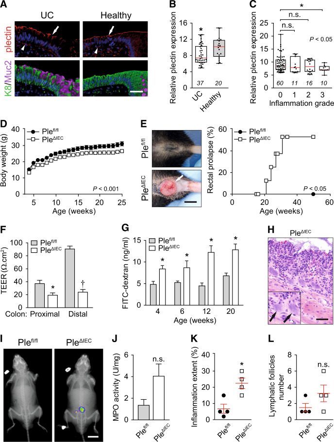
Fig. 1. Loss of plectin is associated with UC in human patients and leads to intestinal epithelial barrier dysfunction with concomitant inflammation in mouse.
A Paraffin-embedded colon sections from UC patients (UC) and healthy controls (healthy) were immunolabeled with antibodies to plectin (red), keratin 8 (K8; green), and mucin 2 (Muc2; magenta). Nuclei were stained with Hoechst (blue). Arrows, apical IEC membrane; arrowheads, basal IEC membrane. Scale bar, 40 μm. B Relative plectin mRNA levels in rectum biopsies collected from healthy controls and patients with active UC. Scattered boxplots show individual data points, median, 25th, and 75th percentile with whiskers reaching the last data point. The numbers of included participants per cohort are indicated in the graph. C Relative plectin mRNA expression in rectum biopsies collected from UC patients clustered based on inflammation scored in H&E-stained rectum sections. Scattered boxplots show individual data points, median, 25th, and 75th percentile with whiskers reaching the last data point. The numbers of included participants per cohort are indicated in the graph. D Bodyweight of Plefl/fl and PleΔIEC mice was monitored for 25 weeks, n = 7. E Representative images of the rectum of 30-week-old Plefl/fl and PleΔIEC mice. Kaplan–Meier graph shows age-related rectal prolapse incidence. F Intestinal transepithelial electrical resistance (TEER) measured ex vivo in both proximal and distal colons of 12-week-old Plefl/fl and PleΔIEC mice, n = 4. G In vivo permeability of mucosa of Plefl/fl and PleΔIEC mice (at the age indicated) measured by monitoring 40-kDa FITC-dextran levels in plasma 4 h after orogastric gavage, n = 3–7. H Representative image of PleΔIEC colon section from 30-week-old PleΔIEC mouse stained with H&E. Arrows, bacterial patches in the mucosa. Scale bar, 50 μm. I In vivo chemiluminescence images of 12-week-old PleΔIEC and Plefl/fl mice injected with myeloperoxidase (MPO) inflammation probe. J MPO activity (a marker of neutrophil infiltration) measured in colon lysates from 12-week-old PleΔIEC and Plefl/fl mice, n = 3. K, L Inflammation extent (percentage) (K) and the number of lymphatic follicles (L) assessed from H&E-stained sections of 12-week-old Plefl/fl and PleΔIEC colons, n = 4. Data are presented as mean ± SEM, n.s. not significant, *P < 0.05, **P < 0.01, †P < 0.001.

