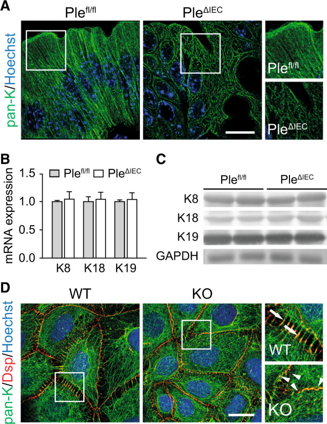
Fig. 4. Plectin organizes KFs in IECs.

A Representative super-resolution STED images of Plefl/fl and PleΔIEC distal colon sections immunolabeled for pan-keratin (pan-K; green) with nuclei stained with Hoechst (blue). Scale bar, 10 μm. Boxed areas show ×1.3 images. B, C Relative mRNA (B) and protein (C) levels of K8, 18, and 19 in Plefl/fl and PleΔIEC distal colon, n = 3–5. Data are presented as mean ± SEM, P > 0.05 by unpaired Student t test. D Representative immunofluorescence images of WT and KO Caco-2 cell monolayer cultures immunolabeled for pan-K (green) and desmoplakin (Dsp; red). Nuclei were stained with Hoechst (blue). Arrows, straight K8 filaments anchored to Dsp-positive desmosomes; arrowheads, tangled K8 filaments. Scale bar, 20 μm. Boxed areas show ×2.5 images.
