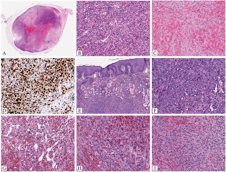
Figure 1. Morphologic spectrum of EH cases with GATA6-FOXO1 fusions.
A-D. Intravascular EH presenting as a 1 cm painful nodule in the subcutis of the right leg of a 42 year-old female (case 1, index case). A. Whole mount showing an intravascular lesion with variegated cellularity and central hemorrhage. B. High power of the increased cellularity component showing well-formed branching vascular channels, surrounded by brisk lympho-plasmacytic infiltrate. C. Central hemorrhagic area is composed of epithelioid endothelial cells arranged in cords, sheets or ill-defined lumina, obscured by abundant extravasated erythrocytes. D. Tumor cells are strongly and diffusely positive for ERG. E-F (case 2, cheek) Cutaneous EH showing a biphasic architectural pattern, with well-defined vascular channels in subepidermal location, while the solid component present in the deeper dermis. G-I. (case 3, dura) EH with focal vasoformative growth composed of sinusoidal, inter-anastomosing pattern (G), and large areas of solid growth (H,I). Tumor cells show abundant eosinophilic cytoplasm and enlarged nuclei with irregular nuclear contours, open chromatin, small but distinct nucleoli, and mild to moderate nuclear pleomorphism.

