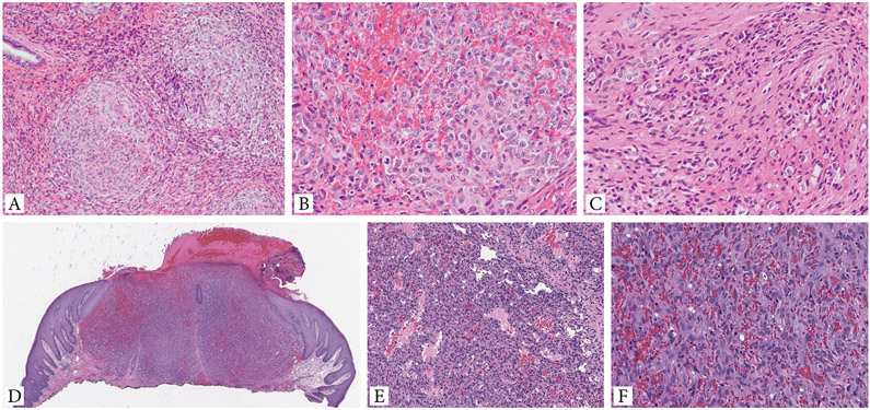
Figure 2. Additional histologic features of EH with GATA6-FOXO1 fusions.
A-C. (case 4) EH involving nasopharyngeal mucosa shows a distinctive multinodular growth (A). Higher power shows solid sheets of epithelioid cells with light eosinophilic cytoplasm and enlarged nuclei with mild atypia, vesicular chromatin and prominent nucleoli. Scattered mitotic figures can be seen, but no necrosis present (B). Extravasated red blood cells are seen intermixed within the solid component. A focal spindle cell component is noted (C). D-F. Cutaneous EH (case 5, back) showing a solid nodular growth, associated with overlying ulceration and delineated by a vague collarette. Tumor displays a biphasic growth, with alternating vasoformative areas composed of sinusoidal and irregular-contour vascular channels (E), and solid components (F). The latter finding reveals sheets of plump ovoid to epithelioid cells with abundant eosinophilic cytoplasm and irregular nuclei with open chromatin and moderate cytologic atypia. No necrosis is identified.

