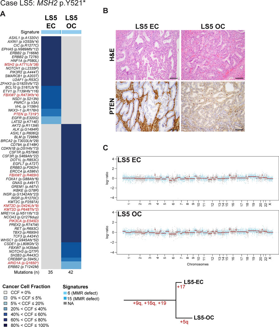
Figure 2: Somatic mutations, mutational signatures, copy number alterations and histologic features of the synchronous endometrial and ovarian cancers from Lynch syndrome case LS5.
(A) Cancer cell fractions of somatic mutations identified in the endometrial cancer (EC) and synchronous ovarian cancer (OC) of case LS5 harboring a germline MSH2 p.Y521* mutation, color coded according to the legend. The EC and OC both display a dominant mutational signature related to DNA mismatch repair defects (Signature 6). Selected pathogenic mutations are highlighted in red font. The total number of somatic mutations identified in the EC and OC are shown below the heatmap. (B) Micrographs of representative hematoxylin and eosin (H&E) and PTEN stained sections of the endometrioid EC and OC (scale bar, 100 μm). Loss of PTEN expression was found only in the EC harboring a PTEN somatic mutation. (C) Chromosome plots of the EC and OC, with the Log2-ratios plotted on the y-axis and the genomic positions on the x-axis (top). Phylogenetic tree based on the copy number alterations depicting the evolution of the EC and OC (bottom). CCF, cancer cell fraction; EC, endometrial carcinoma; OC, ovarian carcinoma.

