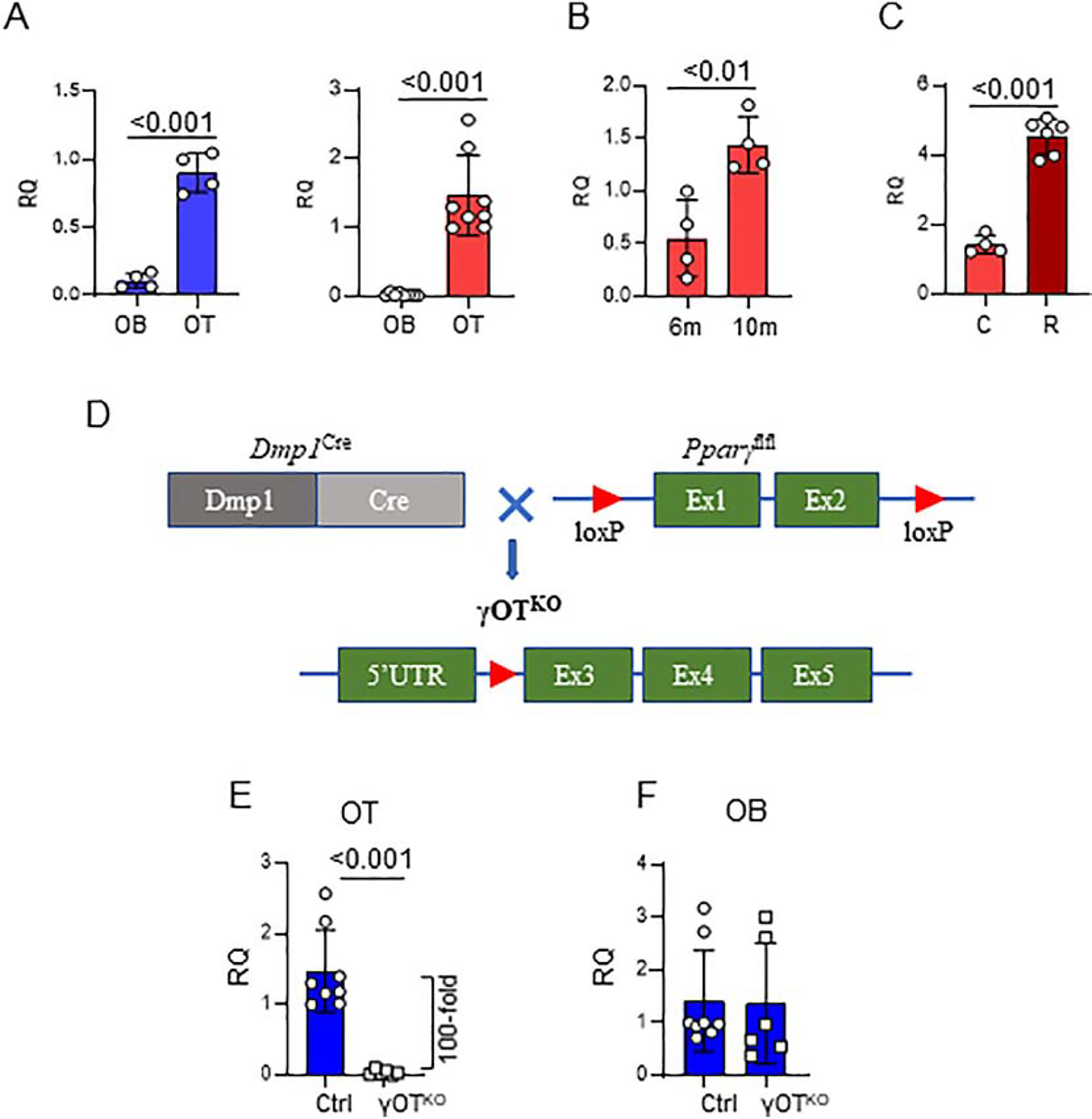
Figure 1.

Development of γOTKO mice. A. Pparγ is highly expressed osteocytes (OT) as compared to osteoblasts (OB). OT and OB were isolated from femora cortical bone of 6.5 – 7 mo old C57BL/6 males (blue) (γOTKO n=4 and Ctrl n=4) and females (red) (γOTKO n=8 and Ctrl n=8) using a method of differential collagen digestion and immediately processed for RNA isolation. B. Pparγ expression in OT increases with aging. OT were isolated as above from femora of 6 mo and 10 mo old C57BL/6 female mice. C. Rosiglitazone treatment increases expression of osteocytic Pparγ. OT were isolated from 10 mo old C57BL/6 female mice treated with 25 mg/kg/d rosiglitazone (R) for 6 weeks (γOTKO n=6 and Ctrl n=4). D. Schematic of γOTKO mice development. γOTKO mice have deleted exon 1 and 2 from Pparγ gene, as a result of crossing Dmp1Cre and Pparγflfl mice. E. Pparγ expression in OT isolated from 6 mo old γOTKO (n=6) and Ctrl (n=8) male mice. F. Pparγ expression in OB isolated from the same mice as in E. Numbers above the horizontal bars indicate p values calculated with parametric unpaired Student t-test. RQ – relative quantity.
