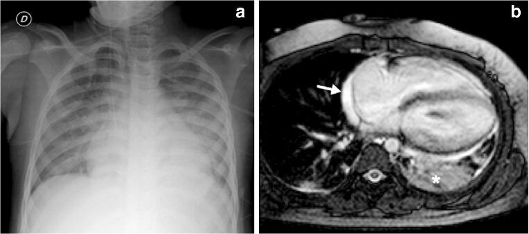
Fig. 2.
Cardiothoracic findings in a 6-year-old boy presenting with impaired biventricular function on echocardiography 6 days after the initial onset of fever, abdominal pain, rash and conjunctivitis. Anti-SARS-CoV-2 (severe acute respiratory syndrome coronavirus 2) total immunoglobulins G, A and M were positive. a Anteroposterior chest radiograph shows cardiomegaly, dilated azygos vein, small left pleural effusion, bilateral airspace consolidation and ground-glass opacities as signs of pulmonary oedema. b Axial balanced turbo field echo cardiac MR image shows consolidation in the left lower lobe (asterisk), small pericardial effusion (arrow) and bilateral small pleural effusion

