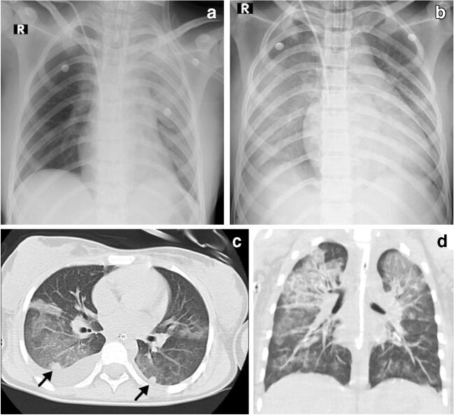
Fig. 3.
Cardiothoracic findings in a 12-year-old girl with fever, abdominal pain, rash, diarrhoea and hypotension who required admission to the intensive care unit. a Initial anteroposterior (AP) radiograph on day 3 reveals focal left upper lobe consolidation and ground-glass opacity. b On day 5 she developed refractory hypoxemia and her AP chest radiograph reveals bilateral diffuse ground-glass opacities, bilateral pleural effusion, focal consolidation in the right upper lobe and interval enlargement of the cardiac silhouette, suggestive of acute respiratory distress syndrome. c, d Axial (c) and coronal (d) images from a chest CT performed the same day as (b) shows bilateral upper lobe ground-glass opacity, septal thickening, right pleural effusion, and nodular consolidation/pulmonary nodules in both lower lobes (arrows)

