Fig. 6.
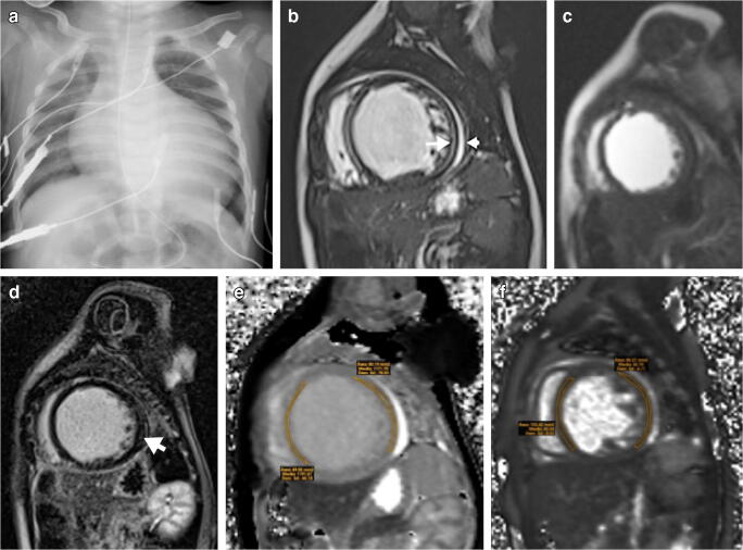
Cardiac failure in a 6-month-old boy with positive SARS-CoV-2 (severe acute respiratory syndrome coronavirus 2) polymerase chain reaction (PCR) test. a Anteroposterior chest radiograph shows cardiomegaly. b–f MRI. Short-axis cardiac MR images show dilated left ventricle with increased signal intensity of the myocardium on balanced fast field echo cine sequence (b) with corresponding early (c) and late (d) gadolinium enhancement on short-axis, saturation-recovery prepared gradient echo (c) and short-axis inversion recovery fast gradient echo (d) images, and increased signal intensity on T1 mapping (e) and T2 mapping (f) (arrows), as well as mild pericardial effusion (arrowhead)

