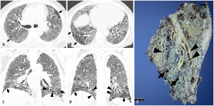
Fig. 1. End-stage pulmonary fibrosis and lung transplantation in a 62-year-old man with usual interstitial pneumonia.
A, B. Lung window images of CT scans obtained at the levels of the right upper lobar bronchus (A) and liver dome (B) show reticular lesions and patchy areas of honeycombing (arrows) in both lungs. C, D. Coronal reformatted images demonstrate areas of honeycombing (arrows) and traction bronchiectasis (arrowheads). E. Gross pathologic specimen of explanted left lung showing areas of honeycombing (arrows) and traction bronchiectasis (arrowheads).

