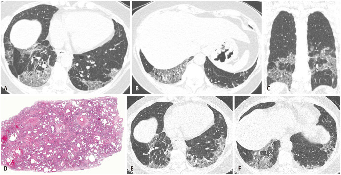
Fig. 2. Fibrotic nonspecific interstitial pneumonia in a 60-year-old woman with Sjogren's syndrome.
A, B. Lung window images of CT scans obtained at the levels of the liver dome (A) and suprahepatic inferior vena cava (B) show mixed areas of ground-glass opacities and reticular lesions with a patchy distribution. C. Coronal reformation image demonstrates lower lung zone predominance of lesions. D. Low-magnification (hematoxylin-eosin staining, × 10) pathological specimen shows chronologically homogeneous (same age) interstitial fibrosis. Uniform thickening of the alveolar septa and the preservation of the lung architecture are noticeable. E, F. Lung window images of CT scans obtained at similar levels to and six years after (A) and (B) without specific treatment show a minimal progression of interstitial lung diseases.

