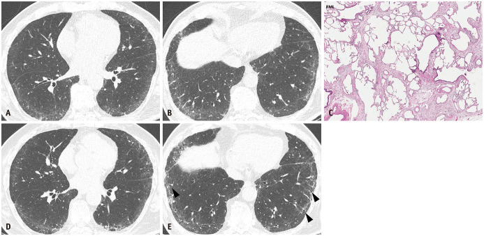
Fig. 3. Fibrotic nonspecific interstitial pneumonia in a 69-year-old man.
A, B. Lung window images of CT scans obtained at the levels of the right inferior pulmonary vein (A) and liver dome (B) show reticular lesions in the subpleural portion of both lungs. C. A high-magnification (hematoxylin-eosin staining, x 100) pathologic specimen obtained from the right middle lobe shows temporally uniform and homogeneous pulmonary fibrosis. D, E. Lung window images of CT scans obtained at similar levels to and four years after (A) and (B) with azathioprine therapy for one and a half years show a minimal progression of pulmonary fibrosis. In addition, the development of traction bronchiolectasis is observable (arrowheads in E).

