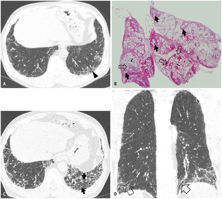
Fig. 4. Pathologic usual interstitial pneumonia manifesting as probable usual interstitial pneumonia pattern on CT in a 62-year-old man.
A. Lung window CT scan image obtained at the level of the liver dome shows reticular lesions in the subpleural portions of the basal lungs. In addition, traction bronchiolectasis (arrowhead) is noticeable. B. Low-magnification (hematoxylin-eosin staining, × 50) pathologic specimen obtained from the right lower lobe discloses interstitial lung disease of temporal heterogeneity composed of areas of microscopic honeycombing (arrows), interstitial fibrosis, and chronic inflammation (open arrows) and normal lungs (small arrows). C. Seven-year follow-up CT scan obtained at a level similar to that in (A) demonstrates the progression of pulmonary fibrosis with new areas of honeycombing (arrows) in the left lower lobe. D. Coronal reformatted image obtained at the same time as (C) shows reticular lesions in the lower lung zones with traction bronchiectasis (open arrows).

