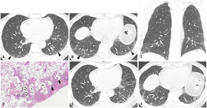
Fig. 5. Pathologic usual interstitial pneumonia manifesting as probable usual interstitial pneumonia pattern on CT in a 61-year-old man.
A, B. Lung window images of CT scans obtained at the levels of the right inferior pulmonary vein (A) and liver dome (B) show reticular lesions in subpleural areas in both lungs, particularly in dependent lungs. Traction bronchiectasis/bronchiolectasis is also observable (arrowheads, visible bronchioles within 2 cm from pleural surfaces). C. Coronal reformatted image demonstrates reticular lesions with subpleural distribution in both lungs. D. High-magnification (hematoxylin-eosin staining, × 200) pathologic specimen obtained from the right lower lobe shows mixed areas of microscopic honeycombing (arrows), interstitial fibrosis, and chronic inflammation (open arrows) and normal lung (small arrows). E, F. Four-year follow-up CT images obtained at similar levels to (A) and (B) and with Pirfenidone (antifibrotic drug) therapy demonstrated a minimal progression of pulmonary fibrosis. In addition, areas of traction bronchiectasis are noticeable (open arrows in E).

