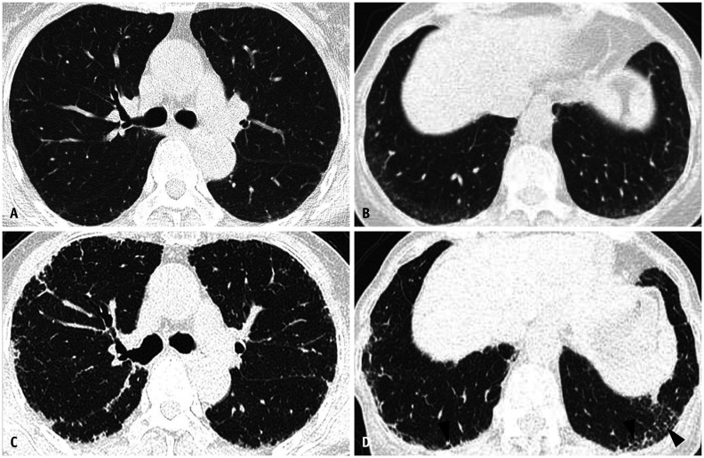
Fig. 7. Evolution of fibrotic interstitial lung abnormality in both lungs during follow-up of eight years in a 70-year-old woman.
A, B. Lung window images of CT scans obtained at the levels of the right upper lobar bronchus (A) and liver dome (B) show fine reticular lesions in the subpleural portion of both lungs. C, D. Eight-year follow-up CT scans obtained at similar levels to (A) and (B) demonstrate a slow progression of pulmonary fibrosis in both lungs; these are interstitial lung diseases. In addition, areas of traction bronchiolectasis are noticeable (arrowheads in D).

