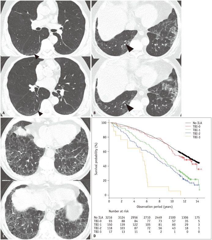
Fig. 8. TBI stratifies prognosis among the subjects with ILA. Reproduced from Hida et al. Eur J Radiol 2020;129:109073 [13].
A. TBI = 1. CT images demonstrate subpleural ground-glass and reticular opacities, indicating the presence of ILA. The dilatation of bronchioles (arrowheads) without obvious architectural distortion in the area of the subpleural opacities of the ILA is shown. B. TBI = 2. CT images demonstrate ground-glass and reticular opacities with subpleural and basilar distributions, indicating the presence of an ILA. Itis characterized by mild bronchiectasis (arrowheads) associated with architectural distortion in the area of subpleural opacities of an ILA. C. TBI = 3. CT images demonstrate ground-glass and reticular opacities with subpleural and basilar distributions, indicating the presence of an ILA. Severe bronchiectasis associated with architectural distortion as well as honeycombing is shown. D. Kaplan-Meier survival curves showing percent survival probability of participants with ILA stratified by TBI = 0, 1, 2, and 3 and those without ILA over years. ILA = interstitial lung abnormality, TBI = traction bronchiectasis index

