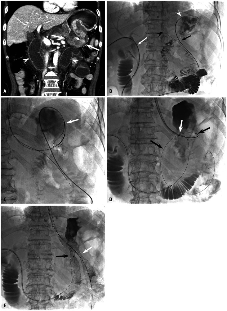
Fig. 1. A 67-year-old man with recurrent obstruction after distal gastrectomy with Billroth II reconstruction.
A. A contrast-enhanced CT shows recurrent tumor at gastrojejunostomy (arrows) with a distended afferent loop (arrowheads). B–E. Photographs obtained during stent placement with a combined transhepatic and peroral approach. B. A 5-French angiographic catheter (Cook Medical) and a 0.035″ guidewire were introduced using transhepatic access (white arrow), manipulated to retrogradely cross the afferent loop obstruction (black arrow), and positioned into the gastric fundus (white arrowhead). The black arrowhead indicates the nasogastric tube. C. The guidewire was captured with a snare catheter (arrow) (ev3 Inc.) and pulled out of the mouth to establish a wire loop. D. A guiding sheath was introduced from the mouth along the wire loop and positioned close to the obstruction. The white arrow indicates the radiopaque marker at the distal end of the guiding sheath. Another angiographic catheter and guidewire assembly was introduced into the guiding sheath and manipulated to cross the efferent loop obstruction (black arrows). E. Two self-expandable stents (S&G BioTech Inc.) were perorally introduced along each guidewire and deployed to cover the efferent (white arrow, 22 mm × 9 cm) and afferent (black arrow, 18 mm × 9 cm) loop obstruction with parallel fashion. Note focal waists at the mid-portion of the stents.

