Figure 2.
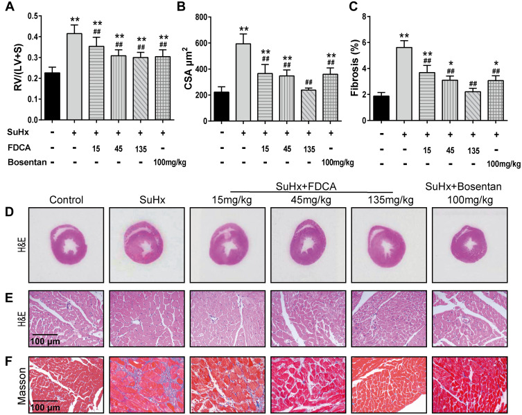
FDCA ameliorated right ventricular (RV) hypertrophy and fibrosis in SuHx-induced rat model of PAH. (A) RV hypertrophy was assessed based on the ratio of the RV weight to the left ventricle and interventricular septum (LV+S) weight (RV/(LV+S)). (B) RV remodeling was also evaluated based on RV cardiomyocyte cross-sectional area (CSA). (C) Degree of fibrosis in RV tissue was calculated based on the percentage of the collagen-positive area. (D) Representative images of H&E staining of full heart cross-sections. (E) Representative images of H&E staining of RV cross-sections. (F) Representative images of Masson’s trichrome staining of RV cross-sections (collagen is stained blue). Data are expressed as mean ± SD (n=6-8). *P<0.05, **P<0.01 vs normoxia control group; ##P<0.01 vs SuHx group.

