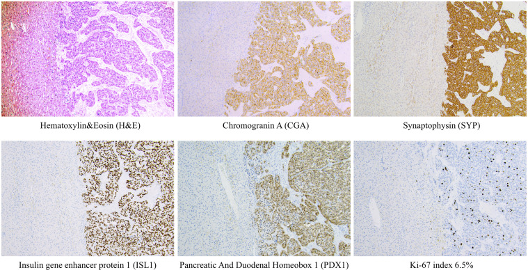
Figure 2.
Examples of routine histological and immunohistochemical features of a metastatic pan-NETs WHO grade 2 (Pan-NET G2) from the Karolinska cohort. Metastatic Pan-NETs tissue is evident to the right, while the left section of each image depicts liver tissue. Note the well-differentiated tumoral growth pattern on routine H&E staining. Tumor cells were diffusely positive for markers of neuroendocrine differentiation (CGA, SYP, ISL1) and displayed stainings indicating a pancreatic origin (ISL1, PDX1). The tumor grade was determined to G2, which in this manuscript would translate to the hypothetical G2a category. This patient had been previously diagnosed with a primary pan-NET (data not shown). All photomicrographs were magnified x100.

