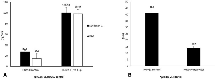
Figure 3.
(A) Glycocalyx shedding in human umbilical vein endothelial cell (HUVEC) monolayers. ELISA kits were used to quantitate syndecan-1 and hyaluronic acid (HLA) levels present in the perfusate collected from HUVEC exposed to hypoxia (Hyp) (1% O2) and epinephrine (epi) perfusion for 90 min. (B) HUVEC glycocalyx thickness measurement. Three-dimensional XYZ image stacks were acquired and processed and analyzed using Volocity cellular imaging and analysis software to assess glycocalyx thickness.

