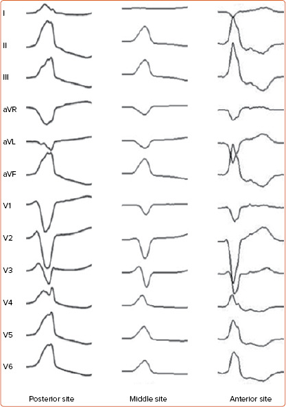
Figure 3: 12-lead ECG of Ventricular Arrhythmias Arising from Right Ventricular Outflow Tract Anterior, Middle and Posterior Sites.

Right ventricular outflow tract (RVOT) ventricular arrhythmias usually present left bundle branch block with late precordial transition and inferior QRS axis. Of note, Q wave amplitude in lead aVR becomes progressively larger moving from anterior to posterior RVOT sites; conversely, Q wave amplitude in lead aVL is smaller in posterior than in anterior RVOT sites. This is due to the caudocephalic spiral orientation of the RVOT, which wraps around the left ventricular outflow tract and is situated progressively anterior and to the left of the aortic root.
