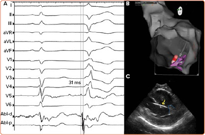
Figure 2: Activation Mapping and Ablation of Premature Ventricular Complexes Originating from the Body of the Postero-medial Papillary Muscle.

A: 12-lead and ablation catheter tracing. The premature complex shows a QS complex in II, III, aVF, R complex from V1 to Ve and RS in lead V6. The ventricular electrogram at earliest activation site preceeded the QRS onset by 31 ms; B: Electroanatomical mapping showing the catheter pointing towards the body of the postero-medial papillary muscle (pink). A clipping plane was applied to the apex of the left ventricle in order to facilitate the view of the postero-medial papillary muscle; C: Ultrasound view of the postero-medial papillary muscle (light blue line) and ablation catheter (yellow arrow) placed at the earliest activation site, through a transmitral approach.
