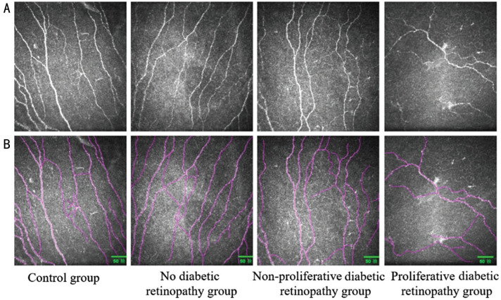
Figure 3. Corneal subbasal nerve plexus analysis.
A: The original image of corneal subbasal nerve plexus morphology; B: Nerves detected with the Image J image analysis software and Neuron J are shown in purple. With the gradual aggravation of diabetic retinopathy, the number and density of corneal nerve fibers decrease, and the nerve fibers becomes deform and irregular.

