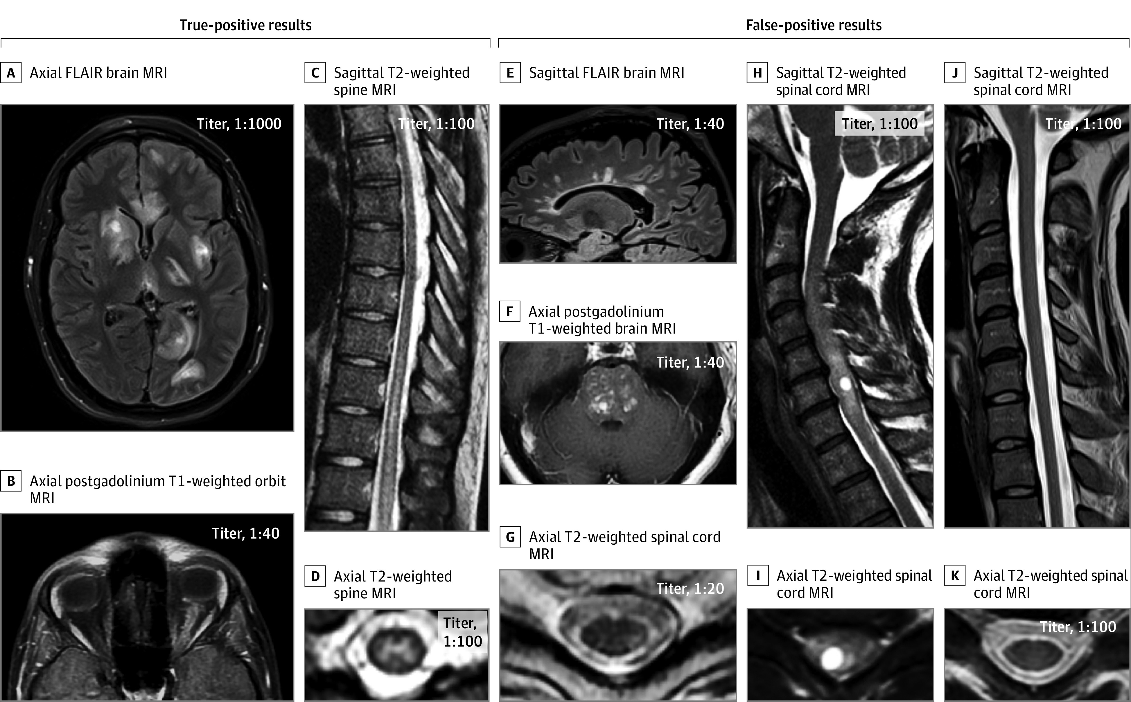
Figure 2. Representative Examples of Magnetic Resonance Imaging (MRI) Findings in True-Positive vs False-Positive Myelin Oligodendrocyte Glycoprotein (MOG)–IgG1 Cases.

True-positive cases (A-D): axial fluid-attenuated inversion recovery (FLAIR) image showing large, multifocal, poorly demarcated lesions on brain MRI in a patient with an acute disseminated encephalomyelitis attack in MOG-IgG1–associated disorder (MOGAD) (A); axial postgadolinium T1-weighted orbit MRI showing longitudinally extensive enhancement of the left optic nerve sheath in a patient with optic neuritis as a manifestation of MOGAD (B); sagittal T2-weighted images showing a longitudinally extensive myelitis lesion along the lower thoracic spinal cord, with predominant involvement of the central gray-matter on axial images in MOGAD (C and D). False-positive cases (E-K): sagittal FLAIR image showing Dawson-finger T2-hyperintense lesions perpendicular to the ventricle, typical of multiple sclerosis (E); axial postgadolinium T1-weighted images showing multiple areas of nodular enhancement in the pons that brainstem biopsy confirmed to be a histiocytic disorder (F); axial T2-weighted image showing a peripheral dorsolateral hyperintense lesion abutting the surface of the spinal cord, in another patient with multiple sclerosis (G); sagittal T2-weighted image showing a faint longitudinally extensive T2-hyperintense lesion accompanied by marked cervical spinal cord swelling with an intralesional cyst (H), also appreciable on axial images (I), which biopsy confirmed to be a glioma; sagittal (J) and axial (K) T2-weighted spinal cord MRI showing normal signal intensity and initial atrophy in a young adult man with X-linked adrenomyeloneuropathy.
